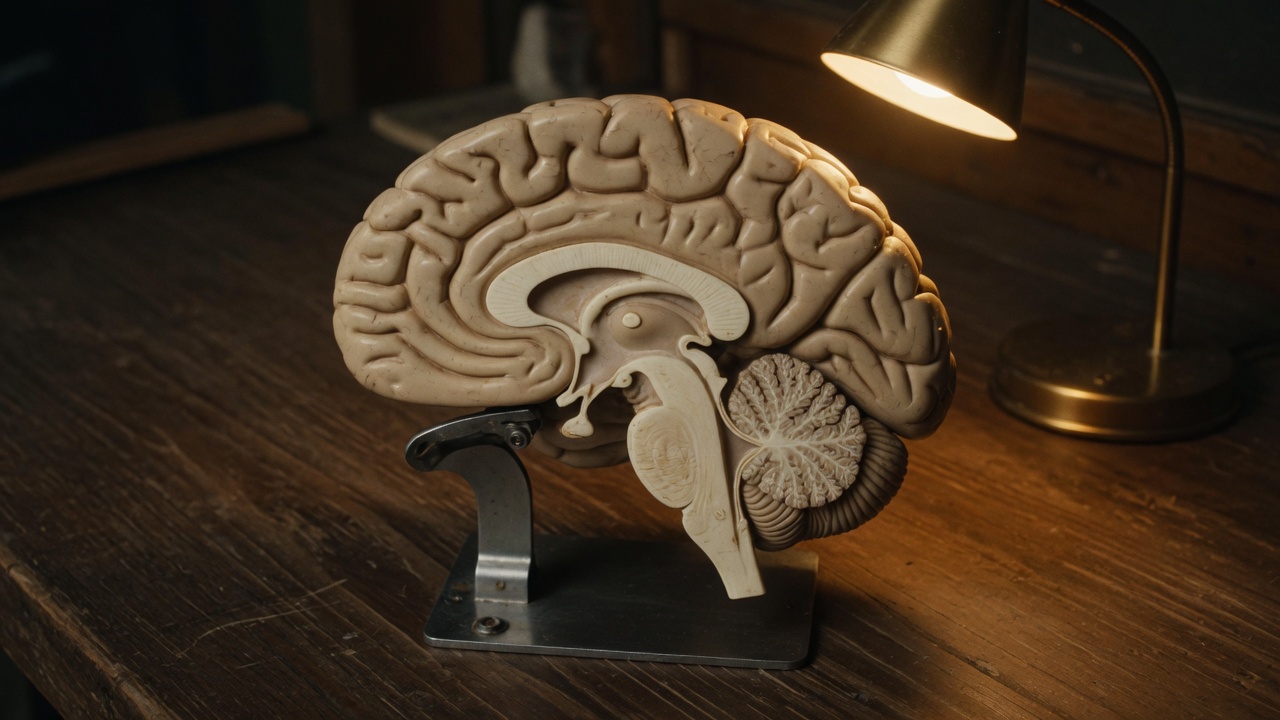
If a person can no longer name objects after a stroke, what has to have been true about the brain?
Some tissue that supported naming was damaged. A stroke is an interruption of blood flow to part of the brain, and it causes a loss of function in the area it hits. When the damage is small, researchers can link the change in behavior to that one area (Spielman et al., 2020).
That idea has a name: . Particular parts of the brain contribute more to particular jobs (Betts et al., 2022). It is not the old idea that one bump on the skull equals one trait. A region can be more involved in a job and still work as part of a network. In the end, all areas of the brain interact to produce our thoughts and behavior (Spielman et al., 2020; Myers & DeWall, 2021).
This lesson gives you a working map. First the whole nervous system, then the brain from the outside in.
How is the nervous system organized?
The nervous system has two main parts. The (CNS) is the brain and the spinal cord. The (PNS) is everything else: the nerves that connect the CNS to the muscles, organs, and senses of the body (Spielman et al., 2020). A nerve is a thick bundle of axons, the long sending fibers of neurons, that carries messages back and forth.
The PNS splits again into two parts.
- The handles things we think of as voluntary. Sensory neurons carry information in from the senses. Motor neurons carry commands out to the skeletal muscles. An easy way to remember the direction: afferent fibers arrive at the CNS, and efferent fibers exit it (Spielman et al., 2020).
- The runs the internal organs and glands, mostly without our control. It works the heart, the smooth muscle of organs like the gut, and glands such as sweat glands (Betts et al., 2022).
The autonomic system has two divisions of its own. The prepares the body for stress and action. The returns the body to routine, everyday operation. They work as a team to keep the body in , a steady balance in things like body temperature (Spielman et al., 2020). Figure 4.1 puts the whole tree on one page.
What does your body do when something startles you?
Picture a hunter who suddenly disturbs a large bear with her cubs. In that instant the sympathetic division takes over. The pupils widen, the heart rate and blood pressure rise, and the liver releases glucose, a sugar the body burns for energy. Adrenaline surges into the bloodstream. This set of changes is the fight-or-flight response. It gives the body quick access to energy so it can fight off a threat or run to safety (Spielman et al., 2020).
Other changes join in. Breathing speeds up to bring in more oxygen, more blood flows to the skeletal muscles, and sweating increases to shed the heat those muscles make. Digestion slows down, so blood is not busy absorbing nutrients when the muscles need oxygen (Betts et al., 2022). Many organs receive nerves from both divisions. The heart is one example: one division speeds it up, and the other slows it down (Betts et al., 2022).
Once the threat is over, the parasympathetic division takes over. Heart rate and blood pressure return to normal, the pupils narrow, and the liver begins to store glucose again as glycogen for later use (Spielman et al., 2020). This is why the parasympathetic division is nicknamed rest and digest (Betts et al., 2022). Some writers now add "fright" and "freeze" to "fight or flight," since not everyone fights or runs when afraid (Betts et al., 2022).
Most of our modern threats are not bears. A speech or a big test can set off the same response, even though there is no physical danger. When stress keeps coming and we can neither fight nor flee, it takes a toll. Research links repeated stress to a higher risk of heart disease and weaker immune function (Spielman et al., 2020). Try Figure 4.2 to see both divisions at work.
Parasympathetic division active: rest and digest
What does the spinal cord do?
The spinal cord connects the brain to the rest of the body. It works like a relay station, but a smart one. Sensory nerves bring messages in, motor nerves send messages out, and messages travel to and from the brain through every segment (Spielman et al., 2020).
Some messages never reach the brain. When you touch a hot stove, the spinal cord starts the pull-away on its own. This automatic response is a , and it saves precious time. The knee jerk is another example (Spielman et al., 2020).
Because every segment passes messages along, an injury to the cord cuts off all the segments below it from the brain. The result is paralysis below the injury. The lower the damage, the fewer functions are lost (Spielman et al., 2020).
How is the brain laid out?
One simple way to divide the brain is into three units: the forebrain, the midbrain, and the hindbrain (NINDS, n.d.). The forebrain is the largest part. It holds the cerebrum and the structures tucked beneath it. The midbrain sits deep in the middle. The hindbrain sits at the back of the head, where the brain meets the spinal cord (Spielman et al., 2020).
When people picture a brain, they picture the cerebrum, the large wrinkled mass on top. Its outer layer is the , from the Latin word for bark. The cortex is only about as thick as a stack of two or three dimes, yet most of the brain's information processing happens there (NINDS, n.d.). It is the "gray matter." Its folds let more of it fit inside the skull. Spread out flat, it would cover roughly one square meter (Betts et al., 2022). Each ridge is a gyrus, and each groove is a sulcus (Spielman et al., 2020).
A deep groove, the longitudinal fissure, splits the cerebrum into left and right hemispheres. Nearly all signals cross over on their way to and from the brain. So the left hemisphere mainly controls the right side of the body, and the right hemisphere controls the left. A stroke in the right hemisphere can weaken the left arm and leg (NINDS, n.d.). The two halves talk through the , a thick band of about 200 million axons (Spielman et al., 2020).
What do the four lobes do?
Each hemisphere is divided into four lobes, using the big grooves as borders (Betts et al., 2022). Figure 4.3 shows them from the side. Each lobe has a main job in the intro map, but each also works with the others.
Frontal lobe
The frontal lobe lies behind the forehead. It is involved in reasoning, planning, movement, emotion, and language (Spielman et al., 2020). At its very front is the prefrontal cortex, tied to judgment, reasoning, and impulse control. Low on the frontal lobe, usually on the left, is Broca's area, which is essential for producing speech. People with damage there can understand language but struggle to produce it (Spielman et al., 2020; Betts et al., 2022).
The strip along the back edge of the frontal lobe is the motor cortex. It helps plan, control, and carry out voluntary movement, like moving your arm or kicking a ball (NINDS, n.d.). Its cells send commands down to the spinal cord to move the skeletal muscles (Betts et al., 2022).
Parietal lobe
Just behind the frontal lobe is the parietal lobe. It processes information from the body's senses. Its front strip, right behind the motor cortex, is the somatosensory cortex. It handles touch, temperature, pain, and the sense of body position (Spielman et al., 2020; Betts et al., 2022). Each part of this strip handles a different part of the body. Parts with more nerves get more room, so the fingers take up far more space than the toes (Spielman et al., 2020).
Temporal lobe
The temporal lobe sits on the side of the head, near the temples. It is linked to hearing, memory, emotion, and parts of language. It holds the auditory cortex, which processes sound. It also holds Wernicke's area, which matters for understanding speech. Damage to Broca's area makes speaking hard. Damage to Wernicke's area leaves a person able to produce fluent words but unable to understand language (Spielman et al., 2020).
Occipital lobe
The occipital lobe is at the very back of the brain. It holds the primary visual cortex, which interprets what the eyes send in (Spielman et al., 2020). Damage to the occipital lobes can cause blindness (NINDS, n.d.).
A. Outside, left side
B. Inside, cut down the middle
| Lobe | Primary job in the intro map |
|---|---|
| frontal | Planning, judgment, voluntary movement (motor cortex), speech production (Broca's area). |
| parietal | Touch and body sense (somatosensory cortex), spatial attention. |
| temporal | Hearing, understanding language (Wernicke's area), memory. |
| occipital | Vision. |
What lies beneath the cortex?
Several structures sit under the cortex, deep inside the forebrain. Figure 4.3B shows where they are.
The is the brain's sensory relay. Every sense except smell passes through it before reaching the cortex (Spielman et al., 2020). It does more than pass messages along. It also helps decide which signals get attention (Betts et al., 2022).
Just below it is the , about the size of a pearl (NINDS, n.d.). It keeps the body in balance by regulating temperature, appetite, and blood pressure. It is in charge of the autonomic nervous system, and it links the nervous system to the hormone system through the pituitary gland (Spielman et al., 2020; Betts et al., 2022).
The limbic system is a group of structures involved in emotion and memory. Along with the hypothalamus, two of its key parts are the hippocampus and the amygdala (Spielman et al., 2020). The is essential for learning and forming new memories. The is involved in emotion and in tying emotional meaning to memories (Spielman et al., 2020). Smell connects directly to the limbic system, which may be why a scent can bring back a feeling so quickly (Spielman et al., 2020).
A famous case shows what the hippocampus does. In 1953, a 27-year-old man known as H. M. had surgery to remove his hippocampus and amygdala to control severe seizures. His seizures eased, but he lost the ability to form many new memories. He could learn a new skill, yet he would not remember learning it (Spielman et al., 2020).
Clusters called the basal ganglia surround the thalamus. They help start and smooth out movements. Parkinson's disease, with its tremors and stiff, shuffling walk, affects these cells (NINDS, n.d.).
What keeps you alive without your thinking about it?
The brainstem connects the brain to the spinal cord. It is made of the midbrain, the pons, and the medulla (Betts et al., 2022).
- The midbrain is the center of the reticular formation, a network that regulates sleep and waking, arousal, and alertness. The midbrain also holds dopamine-making cells that are critical for movement. Their loss is involved in Parkinson's disease (Spielman et al., 2020).
- The pons, from the Latin word for bridge, connects the hindbrain to the rest of the brain. It also helps regulate brain activity during sleep (Spielman et al., 2020).
- The medulla controls automatic functions such as breathing, blood pressure, and heart rate (Spielman et al., 2020).
Behind the brainstem is the , Latin for "little brain." It makes up about 10 percent of the brain's mass (Betts et al., 2022). It takes in messages from the muscles, joints, and inner ear to control balance, coordination, and motor skills. It compares the movement you meant to make with the movement you actually made, then sends corrections (Spielman et al., 2020; Betts et al., 2022). The cerebellum also seems to store memory for how to do things. That may be why H. M. could still learn new skills: his cerebellum was intact (Spielman et al., 2020).
Figure 4.5 lets you turn a simplified brain in 3D. Tap a lobe on the outside, or turn on "Show inside" to find the deep structures.
What happens when the two halves are cut apart?
In some cases of severe epilepsy, surgeons cut the corpus callosum to stop seizures from spreading. The treatment works, but it leaves a person with a "split brain" (Spielman et al., 2020).
Split-brain patients show something striking. If a picture is flashed only to the left side of their vision, that information goes only to the right hemisphere, which is largely nonverbal. The person cannot name the picture. Yet they can draw it with the left hand, which the right hemisphere controls. Once the more verbal left hemisphere sees the drawing, the person can name it (Spielman et al., 2020).
Findings like this led to a popular myth that people are "left-brained" or "right-brained." The real picture is more modest. There is some specialization: language leans left in most people, and some spatial skills lean right. But neither side has a monopoly, and many functions, including language, are spread across both hemispheres (Betts et al., 2022). It is better to ask how the two halves work together (Spielman et al., 2020).
If function can reorganize after injury, what is that capacity called?
Neuroplasticity, often shortened to . It is the nervous system's ability to change and adapt. It can come from experience, from normal development, or from recovery after an injury. It can mean new synapses, pruning of unused ones, changes in glial cells, and even the birth of new neurons. The brain is most plastic when we are young, but it keeps this ability throughout life (Spielman et al., 2020).
This is part of why people can regain skills after a stroke or brain injury, often with intensive therapy (Spielman et al., 2020). It is also a reminder that the map in this lesson is a starting point. Brains share a common layout, but each one is shaped by the life it has lived.
How do scientists see a living brain?
Much of what we know first came from people with brain damage. Today, imaging lets researchers study healthy brains too (Spielman et al., 2020).
- CT (computerized tomography) combines many x-rays into a picture of brain structure. It is often used to find tumors or shrinkage.
- PET (positron emission tomography) follows a mildly radioactive tracer in the blood to map which areas are more active.
- MRI (magnetic resonance imaging) uses a strong magnetic field to make detailed pictures of structure. fMRI (functional MRI) tracks blood flow and oxygen to show activity changing over time.
- EEG (electroencephalography) records the brain's electrical activity through electrodes on the scalp. It gives precise timing, within milliseconds, but little detail about location.
These tools are why this lesson can say "more involved in" instead of guessing (Spielman et al., 2020).
So, back to the person who cannot name objects after a stroke. Some tissue that supported naming, likely in the language areas of the left hemisphere for most people, was damaged. The loss is real, but so is the network around it, and so is plasticity. Later lessons will return to this map when we study sensation, memory, emotion, and stress.
Check yourself
What two structures make up the central nervous system?
The brain and the spinal cord.
Your heart pounds before a big test. Which division of the autonomic nervous system is most active?
The sympathetic division, which drives the fight-or-flight response.
A person can understand speech but struggles to produce it. Which area was most likely damaged?
Broca's area, in the frontal lobe (usually on the left).
Which structure relays every sense except smell?
The thalamus.
H. M. could learn new skills but not new facts. Which structures explain each?
His hippocampus was removed, so he could not form many new memories of facts and events. His cerebellum was intact, so he could still learn skills.
Why can't a split-brain patient name a picture shown only to the left visual field?
The image reaches only the right hemisphere, which is largely nonverbal, and the cut corpus callosum stops it from reaching the left hemisphere's language areas.
Check yourself with a quick quiz
A few questions on this lesson only. Questions you miss come back later. Progress stays in this browser and is shared with the full quiz.
References
- Browne, K., & Schlehlein, H. (2023). 3D reference organ for brain, male (Version 1.3) [3D model]. Human Reference Atlas. Licensed CC BY 4.0. https://doi.org/10.48539/HBM929.XKCL.339
- Betts, J. G., Young, K. A., Wise, J. A., Johnson, E., Poe, B., Kruse, D. H., Korol, O., Johnson, J. E., Womble, M., & DeSaix, P. (2022). Anatomy and physiology 2e. OpenStax. Sections 13.2 The central nervous system and 15.1 Divisions of the autonomic nervous system. https://openstax.org/books/anatomy-and-physiology-2e/pages/13-2-the-central-nervous-system
- Myers, D. G., & DeWall, C. N. (2021). Psychology (13th ed.). Worth.
- National Institute of Neurological Disorders and Stroke. (n.d.). Brain basics: Know your brain. National Institutes of Health. Retrieved September 29, 2026, from https://www.ninds.nih.gov/health-information/public-education/brain-basics/brain-basics-know-your-brain
- Spielman, R. M., Jenkins, W. J., & Lovett, M. D. (2020). Psychology 2e. OpenStax. Sections 3.3 Parts of the nervous system and 3.4 The brain and spinal cord. https://openstax.org/books/psychology-2e/pages/3-4-the-brain-and-spinal-cord
Image and media credits
- Top image. An anatomical brain model on a wooden desk under a lamp. It sets the scene for this lesson on the brain and nervous system. Source: Created for this site with Grok Imagine (xAI), an AI image generator. License: Original image made for mattgaugerx.com.
- Read-aloud audio. Narration of this page. It lets you listen to the lesson instead of reading it. Source: Created for this site: this page's text read by a computer-generated voice (Microsoft text-to-speech, Andrew voice). License: Our own text; the synthetic voice is Microsoft's.
- Brain renderings and 3D brain. The brain pictures in Figures 4.1 and 4.3 and the turnable 3D brain in Figure 4.5, adapted from a 3D reference model: regions merged, simplified, and recolored. They show where each brain region sits, from the outside and from the middle. Source: Kristen Browne and Heidi Schlehlein, 3D Reference Organ for Brain, Male v1.3 (2023), Human Reference Atlas (HuBMAP, NIH), based on the Allen Human Reference Atlas, doi.org/10.48539/HBM929.XKCL.339. License: CC BY 4.0.
- Diagrams and interactive figures. Every diagram and interactive figure on this page apart from the brain renderings credited above. They show the ideas in the text step by step. Source: Created for this site. License: Original to mattgaugerx.com.
- 3D viewer software. The interactive 3D figure on this page runs on these open-source libraries. Source: three.js r169 and the Draco mesh decoder (Google). License: MIT License (three.js); Apache License 2.0 (Draco).